

Quick answer
Gamma rays are suitable for cancer treatment because they are a form of ionizing radiation: extremely high-energy, very short-wavelength photons that penetrate deep into the body and carry enough energy to break the DNA of cancer cells, stopping them from dividing. Because they have no charge and travel in straight lines, beams can be aimed from many angles to meet at a tumor, and treatment is given in small daily doses so that healthy cells have time to repair themselves.
It is a question that appears in school physics textbooks and in the minds of patients preparing for radiotherapy alike: what special property do gamma rays have that makes them suitable for cancer treatment? The answer lies in their remarkable energy, their ability to pass deep into the body and the way they damage the genetic material of rapidly dividing cells.
This guide explains the science in plain language, shows how gamma rays and their close cousins, high-energy X-rays, are used in modern cancer care, and describes how radiation fits alongside surgery and chemotherapy.
What Are Gamma Rays?
Gamma rays are the most energetic form of electromagnetic radiation, sitting beyond X-rays at the far end of the spectrum that also includes radio waves, visible light and ultraviolet rays. They are packets of energy called photons, released from the nucleus of an atom during radioactive decay. Cobalt-60, for example, emits gamma rays with energies of about 1.17 and 1.33 million electron volts (MeV), hundreds of thousands of times more energetic than photons of visible light.
| Property | Gamma rays | Why it matters in cancer treatment |
|---|---|---|
| Energy | Very high, typically in the MeV range for therapy | Enough to ionize atoms and damage DNA |
| Wavelength | Extremely short, smaller than an atom | Passes through skin and tissue to reach deep tumors |
| Electric charge and mass | None | Travels in straight lines, so beams can be aimed precisely |
| Ionizing ability | Strong | Creates free radicals that break DNA strands |
| Source | Radioactive decay, such as cobalt-60 or iridium-192 | Provides a steady, predictable output of radiation |
The Key Property: Ionizing Energy That Damages Cancer Cell DNA
The single most important property of gamma rays is that they are ionizing radiation. When a gamma ray passes through tissue, it knocks electrons out of atoms. This can break DNA directly, but most of the damage happens indirectly: the radiation splits water molecules inside cells into highly reactive free radicals, which then attack the DNA.
A cell with badly damaged DNA, especially with breaks in both strands, cannot divide properly and eventually dies, often when it next tries to divide. Cancer cells divide more often than most normal cells and frequently have faulty DNA repair systems, which makes them more vulnerable. Many healthy cells, by contrast, can repair much of the damage if they are given time between doses.
Deep Penetration and Precise Targeting
Gamma rays and high-energy X-rays penetrate deeply, so they can reach tumors in the lung, pelvis or brain without an incision. Because they travel in straight lines, radiation teams can direct several beams from different angles so that they cross at the tumor. Each beam delivers a modest dose along its path, but where the beams overlap, the tumor receives a high dose.
High-energy photons also have a skin-sparing effect: their maximum dose is deposited a little below the skin surface, which helps protect the skin when deeper tumors are treated.
How Gamma Rays Are Used in Cancer Treatment Today
Cobalt-60 Teletherapy
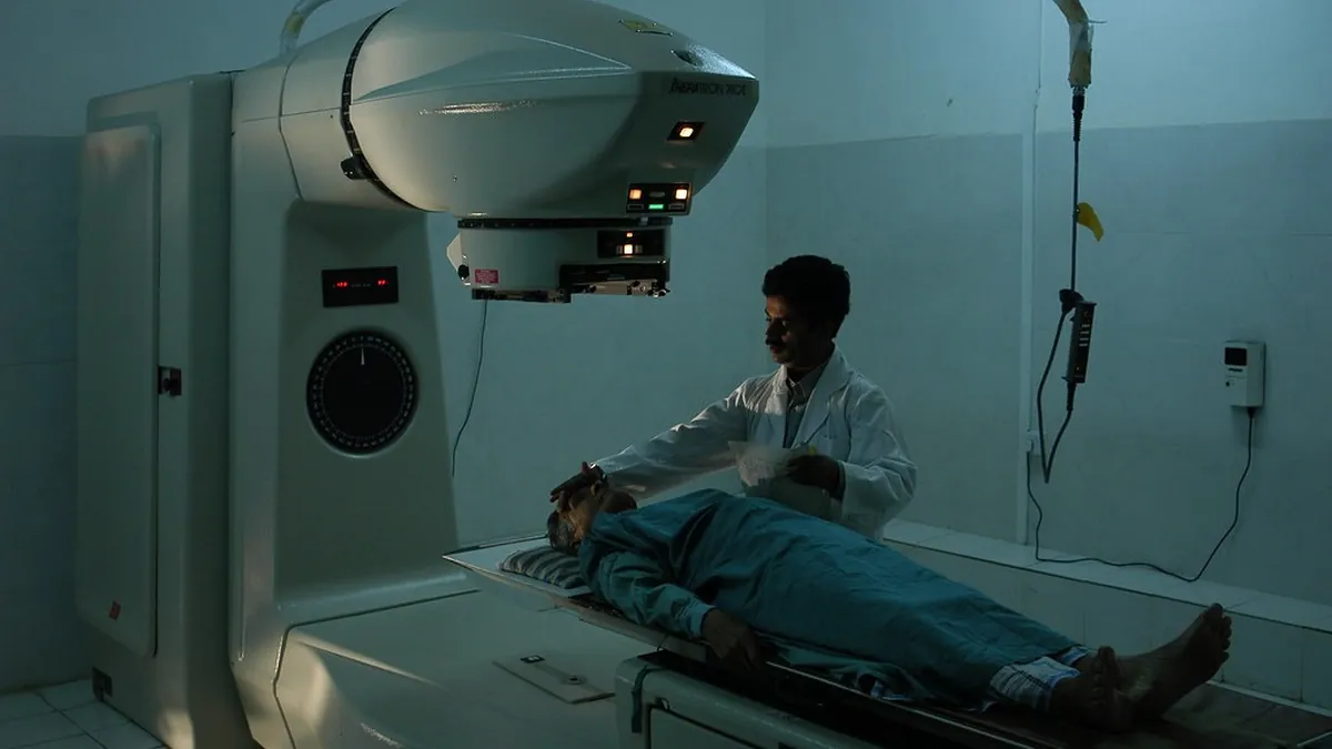
Cobalt-60 machines, first used for patients in the early 1950s, direct a beam of gamma rays from a radioactive source at the tumor. They are robust and relatively simple to operate, and India has developed its own cobalt units, such as the Bhabhatron designed by the Bhabha Atomic Research Centre, to widen access to radiotherapy.
Gamma Knife Radiosurgery
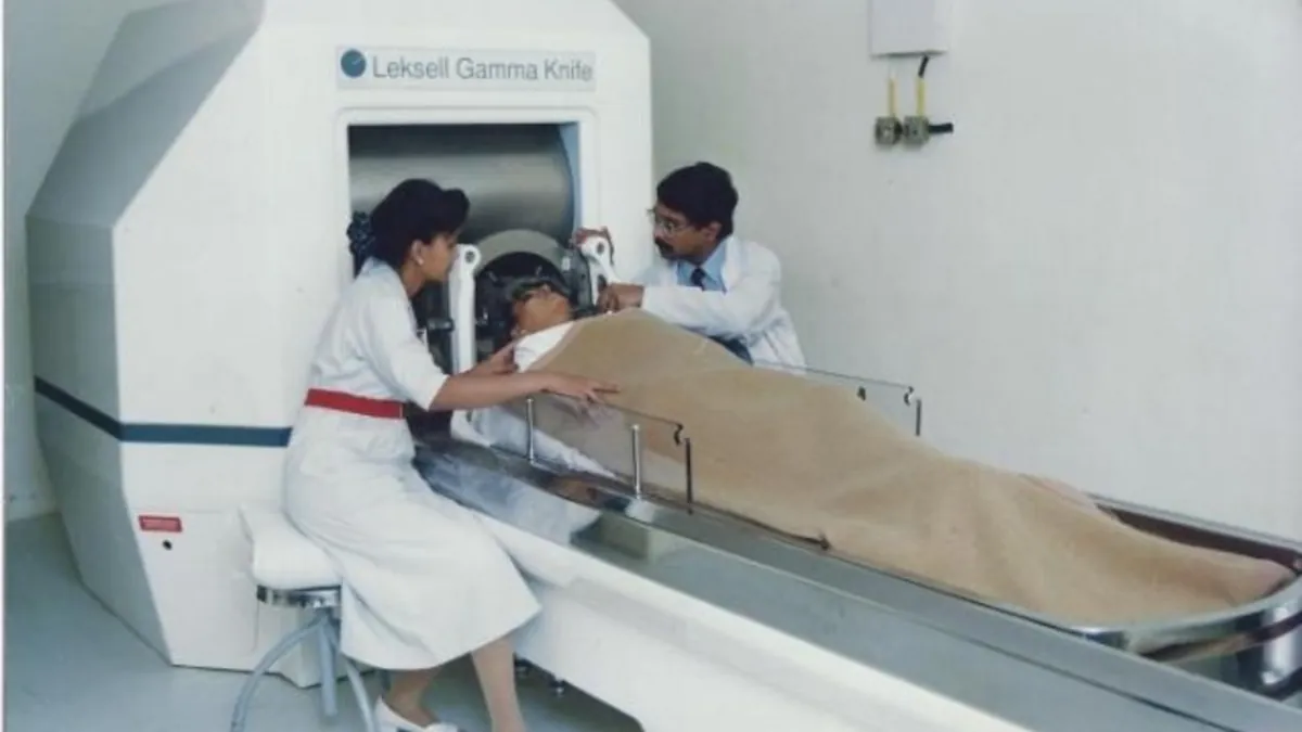
The Gamma Knife focuses around 200 small beams of cobalt-60 gamma rays onto a precise point in the brain. It treats brain metastases, some benign tumors and vascular malformations, usually in a single session without any cut. People who notice the warning signs described in our guide on how to detect a brain tumor at home may, after diagnosis, be assessed for this kind of focused treatment.
Brachytherapy
In brachytherapy, sealed radioactive sources such as iridium-192, which emits gamma rays, are placed inside or right next to a tumor. Because radiation falls off sharply with distance, the tumor receives a very high dose while nearby organs are spared. It is widely used for cervical cancer within gynecologic cancer treatment and for selected head and neck cancers, as well as for prostate and breast cancers.
Linear Accelerators
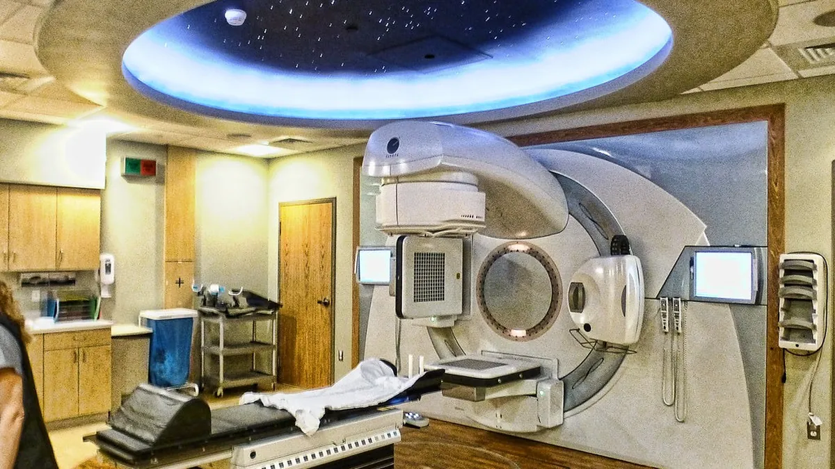
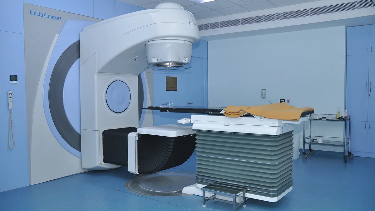
Most modern external beam radiotherapy uses linear accelerators, which produce high-energy X-rays electrically rather than from a radioactive source. These X-rays are photons just like gamma rays and destroy cancer cells in the same way, while allowing advanced techniques such as IMRT, VMAT, image-guided radiotherapy and stereotactic body radiotherapy. The National Cancer Institute’s overview of radiation therapy explains these approaches.
Why Radiation Does Not Simply Destroy Healthy Tissue Too
- Fractionation: the total dose is split into small daily sessions, giving normal cells time to repair between treatments.
- Three-dimensional planning: CT and MRI scans map the tumor and nearby organs so that doses can be calculated precisely.
- Beam shaping: devices called multileaf collimators shape each beam to match the outline of the tumor.
- Image guidance: scans taken before each session confirm that the target is in exactly the right position.
- Dose limits: strict limits protect sensitive organs such as the spinal cord, heart, lungs and bowel.
Common Side Effects of Radiation Therapy
Side effects depend mainly on the area treated and usually build up gradually during the course:
- Tiredness, and skin redness or dryness in the treated area
- A sore mouth and difficulty swallowing when the head and neck are treated
- Cough or breathlessness with radiation to the chest
- Loose motions or bladder irritation with pelvic radiation
Most early effects settle within weeks of finishing treatment. External beam radiation does not make you radioactive, so it is safe to be close to family and children.
How Radiation Works Alongside Surgery
- Before surgery: chemoradiation shrinks tumors and lowers the risk of recurrence before rectal cancer surgery and as part of esophageal cancer treatment.
- After surgery: radiotherapy after breast-conserving surgery is a standard part of breast cancer treatment, and it may also be advised when surgical margins are close.
- Instead of surgery: for small early lung cancers in patients who cannot undergo an operation, stereotactic radiotherapy is an effective alternative, while fit patients are usually offered lung cancer surgery.
Deciding the right order and combination is a key part of a personalized cancer treatment plan, and the same tumor board discussion also decides whether chemotherapy after cancer surgery is needed.
Summary: The Special Properties That Make Gamma Rays Effective
- Very high energy that ionizes atoms and damages cancer cell DNA
- An extremely short wavelength that penetrates deep into the body
- No electric charge, so beams travel straight and can be aimed precisely
- Steady emission from radioactive sources, allowing controlled dosing
Combined with careful planning and fractionation, these properties allow gamma rays and high-energy X-rays to destroy cancer cells while keeping harm to healthy tissue as low as possible.
Frequently Asked Questions
Gamma rays carry very high energy and are ionizing, so they can damage the DNA of cancer cells and stop them from dividing. They also penetrate deep into the body and travel in straight lines, which allows beams to be aimed precisely at tumors.
Cancer cells divide rapidly and are often less able to repair DNA damage. Normal cells repair much of the damage between treatment sessions, which is why radiation is usually given in small daily doses called fractions.
Both are high-energy photons with the same basic effect on cells. Gamma rays come from radioactive decay, such as cobalt-60, while therapeutic X-rays are produced by linear accelerators. Most modern external beam radiotherapy uses high-energy X-rays.
Gamma Knife radiosurgery focuses around 200 small beams of gamma rays from cobalt-60 sources onto a precise target in the brain. It is used for brain metastases, some benign tumors and vascular malformations, often in a single session without an incision.
No. External beam radiation from a machine does not make you radioactive, so you can safely be around family and children. Only some forms of internal radiation, such as permanent seed implants, need short-term precautions, which your team will explain.
Sometimes. For certain early cancers, or for patients who cannot undergo an operation, radiation can be an effective alternative. For many others, surgery and radiation work best together, and a multidisciplinary tumor board decides the right combination.
Medical disclaimer: This article is for general education and does not replace a personal consultation. Cancer care is individual, so please discuss your reports, symptoms and treatment options with a qualified specialist before making any decision.
Image credits: Linear Accelerator - Elekta Compact model by Narenfox, CC BY-SA 4.0 · Dr. B. K. Misra performing Stereotactic Gamma Radiosurgery by Vm1207, CC BY-SA 4.0 · 05810030 by IAEA Imagebank, CC BY-SA 2.0 · radiation scanner by Muffet, CC BY 2.0